Introduction
I would like to start a new subseries in which I start describing different concepts regarding the functioning of the brain, its biology and chemistry.
This series will have an erratic schedule but it should provide a general overview of our brain’s inner workings. These posts are out a series of lectures by Prof. Jan Schnupp, and I want to make sure he is properly quoted and credited. Many parts are his lectures verbatim. However, for any errors, mistakes or inaccuracies in anything I will write in here, or in the next posts, I take all the credit. I probably misunderstood him or made it up.
The three main brain regions
Hindbrain, midbrain and forebrain
The brain grows out of the spinal cord. The spinal cord comes first, both in evolution and in embryology. The spinal cord is actually the first organ that our body ever grows. On top of that, our body grows a brain stem. The brainstem is one of the most important parts of the entire central nervous system, because it connects the brain to the spinal cord and coordinates many vital functions, such as breathing and heartbeat.
And out of the brain stem, at the back, there is the cerebellum. The cerebellum is very important for coordinating movements. Nowadays, people think it may actually do a lot more than that. But that certainly seems to be the most obvious thing. People who are very drunk and staggering and almost falling over behave very much like somebody who's got a big problem with their cerebellum and that's because they just don't have the sort of coordination that the cerebellum gives you. The brainstem (most of it) and the cerebellum are often called the hindbrain.
Image by BruceBlaus - Own work, CC BY-SA 4.0
https://commons.wikimedia.org/w/index.php?curid=47242989
The brain stem and the cerebellum are linked by the midbrain. Normally in the brain, you wouldn't see the midbrain. It's kind of stuck in here, not visible from the outside.
The third major brain region, after the hindbrain and the midbrain is the forebrain. The midbrain links the hindbrain to the forebrain.
The forebrain, the largest brain region, is also divided in three major regions: the thalamus, the hypothalamus and the cerebrum.
The Forebrain
The thalamus in the forebrain is like a relay center that basically connects the large part that most people think of as the brain, which is the cerebral cortex, to the rest of the brain.
So you can think of this almost like a sort of a hierarchy. The thing at the top is the cerebral cortex. Cortex really just means shell or rind. So the rind sits on top of this structure and is linked through the thalamus (the red part in the picture below).
The thalamus can gate activity, can allow information to flow up into the cortex or not. And you can “kind of” switch the cortex off when you go to sleep. Switch it off is actually a highly inaccurate way of putting it, but we'll have more to say about that later.
So that's the rough structure. We've got the cortex at the top. That's what usually people think of when they talk about the brain, the cortex. Linked by the midbrain to the brainstem and cerebellum at the back.
In here we will actually not say much about the cerebellum. Cerebellum is very important for medical doctors to know about because sometimes you get people with injuries there. And then they've got very characteristic motor deficits. But people don't generally think it's the seat of the soul or of things like addiction, love, reasoning, and most of the interesting problems in neuroscience.
The Cortex
The cortex looks like a convoluted mess in a way. But there are very well defined parts to it and we will be able to see that a bit more clearly later.
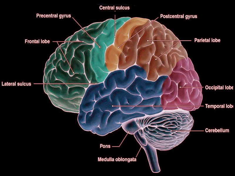
Perhaps the most obvious is that there's a deep fissure running in the middle, the cerebral fissure that splits the brain in two hemispheres. These fissures, if the fissures aren't terribly deep, are called sulci. This is just a Latin word for them. And the ridges between these little fissures are called the gyri.
So we have got gyri and sulci. And we have lots of them and in no two brains the patterns of gyri and sulci are exactly identical.
But they're nevertheless very similar from one person to the next. One thing that you can find in pretty much everybody is one big sulcus that runs along the side.
And that cuts the frontal cortex off the parietal cortex. So we have got frontal and parietal and, at the back of the brain we have occipital. Finally, we also have the temporal cortex, and these are the four different lobes of the cortex. And they do different bits. Each of them have different roles to play. If we were to open one of these things up, if we were to cut into the cortex, then we would see that the cortex is really only about two millimeters deep in humans.
And it's made out of so-called grey matter. It's not really that grey, it's mostly pinkish actually if it's sort of fresh and alive. And then below it is the white matter. The grey matter is full of little nerve cells and if you look at them under the microscope, they look a bit like this.
This is actually a section cut through the cerebral cortex, which is stained with a special stain called a Golgi stain, which will fill individual nerve cells with silver. Little silver granules that form inside them and that will make these stand out. But the funny thing about the Golgi stain, for reasons that nobody really understands, is that it stains only a tiny fraction of all the nerve cells.
Because if you stained all of them, all you'd see is black. They're so densely packed that you wouldn't see any pattern. But it's been very useful for actually seeing individual nerve cells. The picture shows a drawing of a Purkinje cell in the cerebellum cortex done by Santiago Ramón y Cajal, clearly demonstrating the power of Golgi's staining method to reveal fine detail
So each of these is only about 20, 30 microns across. A micron is a millionth of a meter. Millimeters are thousands, microns is a millionth of a meter. So about 20 microns is how big they are.
In the whole of the brain, we have tens of billions of these individual neurons that all have funny shapes like this. The things that are growing out of them are called the dendrites and the axons. They form a dense web of connections. So we have these neuron cell bodies concentrated in the grey matter.
And then we have these white matter tracts. And these white matter tracts are really just cables that grow out of these nerve cells, which are known as axons in the proper nomenclature. Lots and lots of axons. So it's basically as if you had lots and lots of little neurons connected through lots of cables that run through the white matter tract.
If we look at the spinal cord, there too we have grey matter, where we have all the cell bodies, and white matter, where we have lots and lots of axons connecting these things. Now, the spinal cord came first in evolution.
What are the first things that a very primitive animal that can't really do very much other than swim about in a big pond with a flagellum or something needs to do? What it needs to do is just, if something pokes it as a nasty stimulus of some kind, have a reflex that makes it move in a different direction.
And really these sort of very simple primitive reflex arcs are still very much in our spinal cord. So we have neurons that are so-called afferent neurons, because they bring information in.
They come from all over our skin. They connect into the white matter tract of the spinal cord. They connect to cells here in the grey matter. And they allow a flow of information that brings information in, like some painful stimulus, when we touch a hot plate.
The information from the painful stimulus comes into the back, travels into the white matter and connects onto the grey matter neurons. In the grey matter we then have motor neurons that will activate our muscles and cause us to withdraw. So we just have a sort of reflex arc set up. Now the interesting thing about the spinal cord is that for some reasons all of the sensory information seems to be coming in at the back and all the motor information seems to be coming out at the front.
We have so-called dorsal roots, dorsal meaning related to the back, and ventral roots, related to the ventral meaning the belly, coming out at the front. So information goes in at the back and motor commands come out at the front. And at the end of the day, that's what our brains are for. They are collecting sensory information and then we decide what to do about it and we generate movements. And to an extent, the organisation, even of the cerebral cortex, which evolved much, much, much later, still has this sort of front-back difference about it.
There is a lot of sensory processing. Taking information in, for example touch information on the so-called post-central gyrus, visual information in the occipital cortex, auditory information here in the temporal lobes, all of them get combined to help us organise, orient ourself in space, direct our attention to different parts in space and so on.
And then in the pre-central gyrus, we have motor cells that will coordinate our movements.
Image courtesy Dr Chaigasame
The Frontal Cortex
The frontal cortex has to do with working memory, decision-making and things like that. And also so-called limbic things. When neuroscientists talk about something limbic, they really mean something that has to do with emotion.
The word limbic actually has the same root as the word limbo in Latin. Limbo is really like the purgatory. It's somewhere where you're not quite in heaven and you're not quite in hell. It really means originally border-reach.
And the thing about the limbic structures is that they are all arranged around the edge of the cortex. And they really have to do with emotional processing. The orbital frontal cortex, for example, has much to do about how appetising you find something and so on.
There are exceptions to this rule that sensory information comes in at the back and then decision-making and motor programming comes out at the front. The exception is smell, which goes straight into the frontal part. Smell is weird.
Now, all of this is made out of neurons, but not just neurons. There's also other cells in there. Of course, we need blood supply, so we need to have cells that make blood vessels and blood cells themselves. And we also have cells that will form little skins, protective skins and stuff.
But another important class of cells is the so-called glial cells, from the Latin word for glue, which is just simply cells that are not neurons. They don't seem to be doing any information processing, but they do a variety of things that help keep neurons healthy and happy. And they've got a variety of roles.
Only about one in five cells in a brain is a neuron, there are actually more glial cells than there are neurons.
This concludes the rough description of the physical brain. Next time we will discuss how neurons communicate.
See also:
https://byjus.com/neet/difference-between-forebrain-midbrain-and-hindbrain/
https://www.brainline.org/tbi-basics/interactive-brain
Next post: Neurons and Basic Chemistry


Really interesting and I especially appreciated the detailed and at the same time engaging way of explaining it. By the way, I was looking for books or something to read on the topic, and your issue came at the right time, thanks!